An Efficient Automated System for Detection of Diabetic Retinopathy from Fundus Images Using Support Vector Machine and Bayesian Classifiers
US$34.62
10000 in stock
SupportDescription
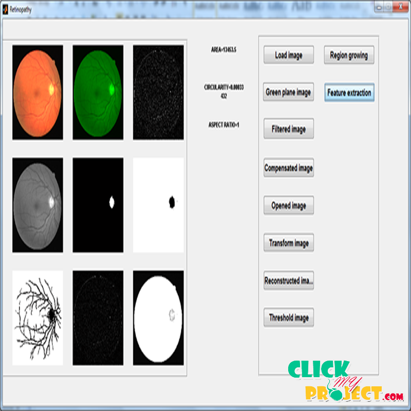
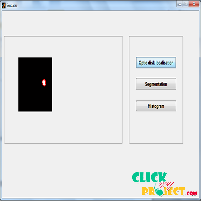
Early diagnosis of diabetes and treatment can prevent vision loss for patients and the preliminary signs of diabetic retinopathy includes micro aneurysms and exudate. The input image is represented in the green plane image in order to reduce the effect of intensity variation. Then the image is filtered using median filter for the removal of noise. After that the image is normalised to get compensated image. Then the transformed image is obtained by the difference between the input image and opened image. In the reconstruction process the geodesic dilation is done . After that the region growing process is done based on image segmentation to partition the image. Then the optic disk is located and segmented. Color histogram technique is used to plot the graph. Finally, we are classifying whether it is exudate or not exudate by using SVM classifier. The preliminary signs of diabetic retinopathy include micro aneurysms, haemorrhages and exudates. Early diagnosis and timely treatment can prevent vision loss in patients with long term diabetes. In this paper we used two algorithm based on filtering operations, morphological transformation and region growing method to extract features for detection of micro aneurysms, haemorrhage and non linear diffusion segmentation followed by colour histogram based clustering techniques is used to differentiate exudates or not exudate. Based on the features obtained, each image is classified as normal or abnormal with Support Vector Machine, Bayesian Network. Classification rate of 95% is obtained with SVM and 90% with Bayesian classifier.


