3D Brain Atlas Reconstruction Using Deformable Medical Image Registration Application to Deep Brain Stimulation Surgery
US$52.63
10000 in stock
SupportDescription
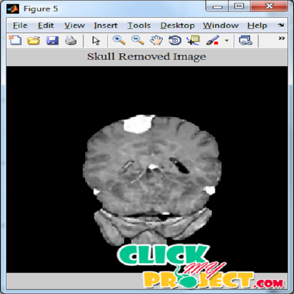
In this proposed work, The brain tumor tissue detection allows to localize a mass of abnormal cells in a slice of Magnetic Resonance (MR). The automatization of this process is useful for post processing of the extracted region of interest like the tumor segmentation. In order to detect this abnormal growth of tissue in an image, this paper presents a novel scheme which uses a two-step procedure; the k-means method and the Hierarchical Centroid Shape Descriptor (HCSD). The clustering stage is applied to discriminate structures based on pixel intensity while the HCSD allow to select only those having a specific shape. A bounding box is then automatically placed to delineate the region in which the tumor was found. Compared to the tumor delineation performed by an expert, a similarity measure of 91% was reached by using the Dice coefficient. The tests were carried out on 254 T1-weighted MRI images of 14 patients with brain tumors. The segmentation in medical images were more helpful for the identification the diseased portion in the images. The preprocessing step is the first step of the segmentation process. The images were filtered using median filter which smoothens the overall image and makes that the intensity all over the image is normalized. Median filter is employed to smoothen the image and to filter unwanted pixels in the image. Median filter parses through the image pixel by pixel, replacing each pixel with the median of neighboring pixels thereby providing normalized intensity in the overall image. The noises in the images were replaced by the above process. The skull regions captured along with the brain region in the images were different intensity and in a large area. The skull regions were identified and removed based on the morphological operations. The morphological operations employed were open operation and close operation. The structuring element is a small binary image, i.e. a small matrix of pixels, each with a value of zero or one: The matrix dimensions specify the size of the structuring element. The pattern of ones and zeros specifies the shape of the structuring element. The closing of an image by a structuring element s is a dilation followed by an erosion. The opening of an image by a structuring element is an erosion followed by a dilation. The tumor portion obtained using thresholding process is considered as the initial portion of the segmentation process. The images pixels surrounding the tumor portion that were having similar values nearby the input region is grouped. Contours were extracted around the detected regions. The process is repeated till the boundary the detection region is within the input region. The final result obtained is the segmented tumor portion. The performance of the process is measured based on the performance metrics like TP, FP, TN, FN, Accuracy, Precision and Recall.
Only logged in customers who have purchased this product may leave a review.


Reviews
There are no reviews yet.