Segmentation-Driven Image Registration-Application to 4D DCE-MRI Recordingsof the Moving Kidneys
₹3,000.00
9999 in stock
SupportDescription
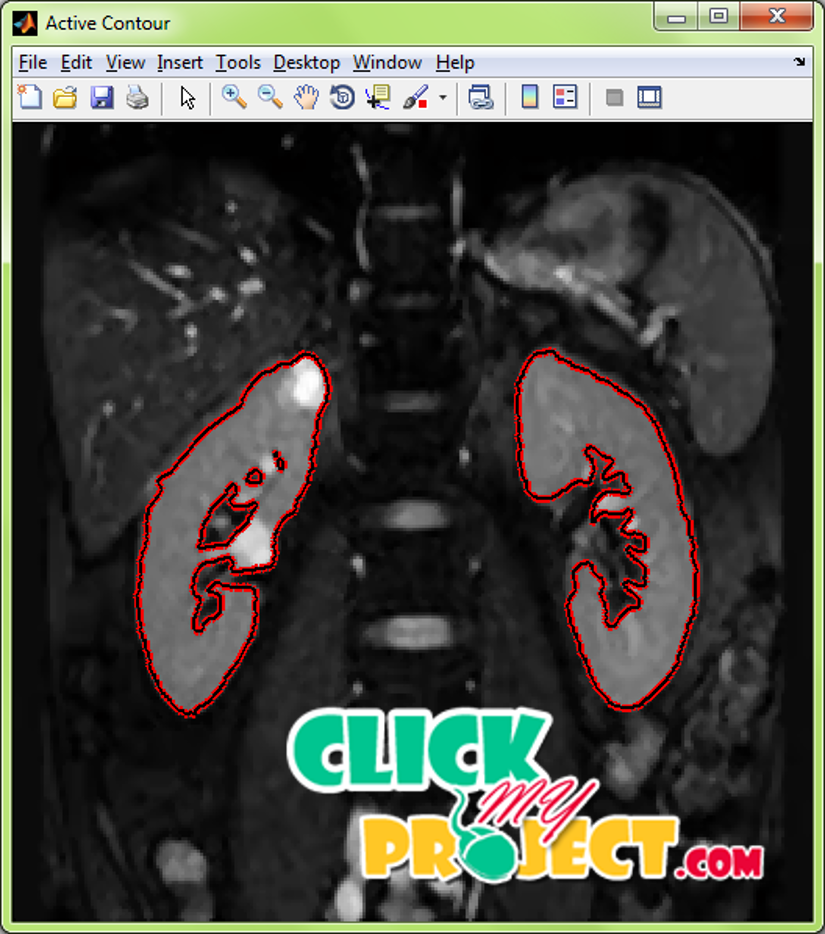
The segmentation of the image refers to extracting the needed region from the image based on some specified methodologies. The segmentation methodologies are commonly based on thresholding (i.e.) the regions within the particular range or pixels that are having particular value. Edge detection, Contour extraction, Clustering are all segmentation methodologies. Edge detection identifies the edge points around the needed objects. Contour extraction refers to outlining the segmented portion from the image. Clustering refers to producing separate identifications (i.e.) color, contour differences for the regions in the image that are similar in intensity or similar in color. Abdominal MRI images has kidney along with other abdominal parts in the image. The segmentation of kidney alone from the MRI images will be useful for the identification of the defects in kidneys. The 2D MRI scan of the abdominal regions were taken. Basic preprocessing steps like gray conversion, resizing and smoothening using filters were applied. These process makes the image pixels to have same sequence of values. The preprocessed images were then segmented by registration process. Several images containing kidney located in different positions were used for training process. The training process registers the shape of the kidney in different positions. For different positions and intensity of the kidney the positions and coordinates of the kidney location in the images were identified. The kidney in the test image is segmented by comparing the shapes in the image with the registered shapes in the dataset. For matching of the kidney the mean value of the images were employed. The calculation of the mean value of the images will give the informations regarding the intensity of the images. The matching process compares the intensity values and based on the needed kidney portion is selected. The selected kidney portion is then extracted from the image. This process is done with the help of Active Contour method. The shape in the dataset that is matching with the input test image is identified with the help of Mahalanobis distance. The distance value will be minimum for the most matching kidney portion in the image. The disease in the kidney images were identified based on the features extracted for the images based on the statistical parameters. The extracted parameters for the process were then classified based on Adaptive Neuro Fuzzy Interface System. The performance of the segmentation process is measured.


