Methods for Nuclei Detection Segmentation and Classification in Digital Histopathology A Review Current Status and Future Potential
₹3,000.00
10000 in stock
SupportDescription
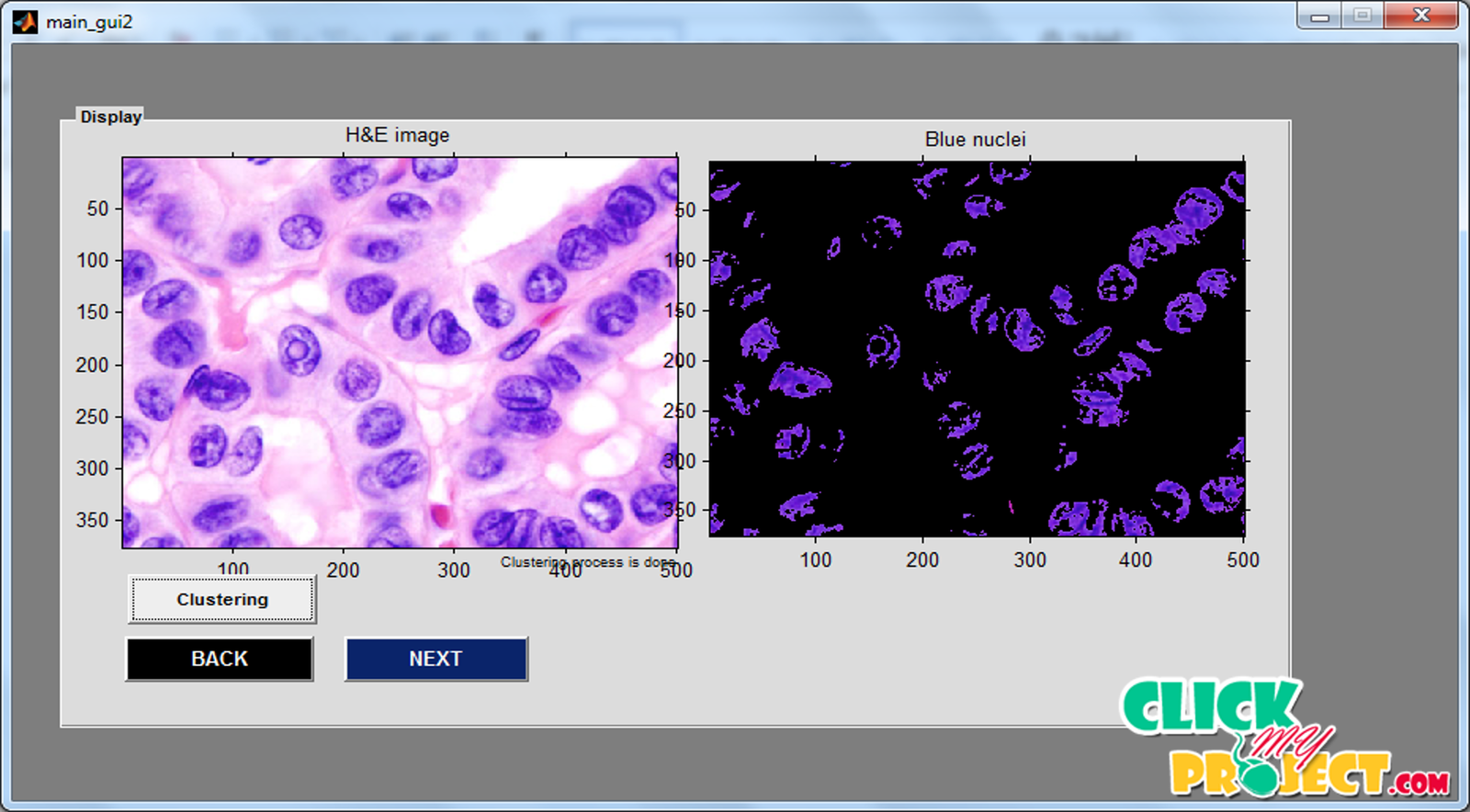
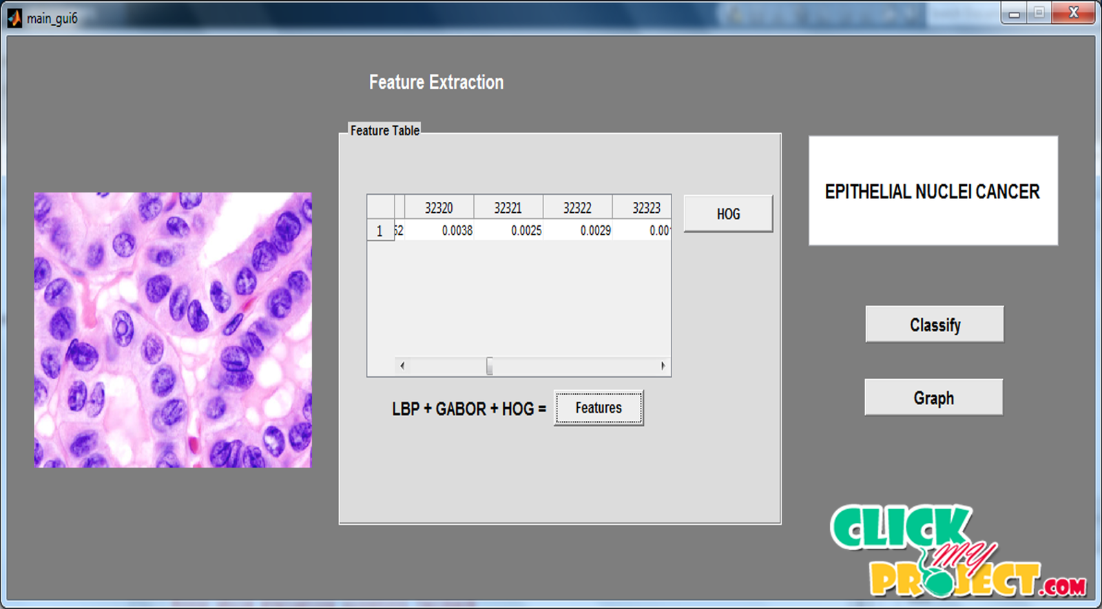
In this method, computerized methods have been rapidly evolving in the area of digital pathology, with growing applications related to nuclei detection, segmentation, and classification. In cancer research, these approaches have played, and will continue to play a major role in minimizing human intervention, consolidating pertinent second opinions, and providing traceable clinical information by this method. To overcome the defect of the cancer cell and abnormality of the cell detection, the segmentation is applicable to detect the cell. Digital pathology represents one of the major evolutions in modern medicine. Pathological examinations constitute the gold standard in many medical protocols, and also play a critical and legal role in the diagnosis process. In the conventional cancer diagnosis, pathologists analyze biopsies to make diagnostic and prognostic assessments, mainly based on the cell morphology and architecture distribution. Recently, computerized methods have been rapidly evolving in the area of digital pathology, with growing applications related to nuclei detection, segmentation, and classification. Each hidden node represents a cluster used as a template to provide faster and more accurate nuclei detection and segmentation. In this paper, we present a fully automated method for annotating fluorescent co focal microscopy images in highly complex conditions. The second layer is comprised of two probabilistic classifiers, responsible for determining how many components may constitute each segmented region.


