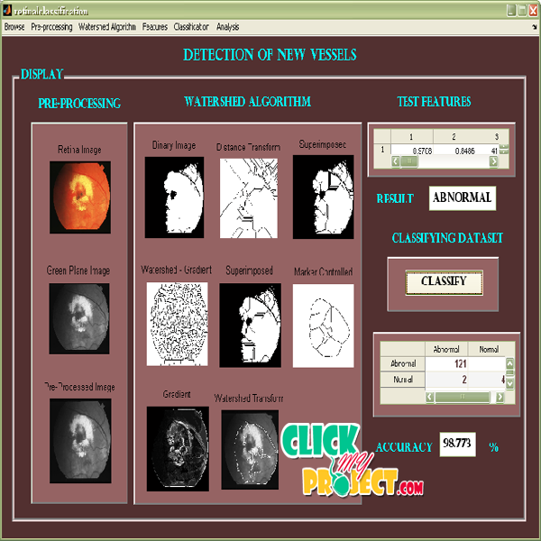
DETECTION OF NEW VESSELS ON THE OPTIC DISC USING RETINAL PHOTOGRAPHS
Rs2,500.00
10000 in stock
SupportDescription
There are two main mechanisms by which vision is lost due to diabetic retinopathy: macular oedema and proliferative retinopathy. Macular oedema is the accumulation of fluid in the macula. Although it cannot be seen directly on monoscopic retinal photographs, its presence may be inferred by surrogate indicators such as exudates. Proliferative retinopathy is the more serious condition as it involves the development of new vessels which are prone to bleed, leading to vitreous haemorrhage, fibrosis, and ultimately retinal detachment. In this paper a method for detecting standard screening photographs which show new vessels on the optic disc is described and evaluated. Of all the features of proliferative retinopathy, newvessels at the disc carry theworst prognosis and detection of these is most likely to add value to an automated grading system. Fifteen feature parameters, associated with shape, position, orientation, brightness, contrast and line density are calculated for each candidate segment. Based on these features, each segment is categorized as normal or abnormal using a support vector machine (SVM) classifier.
Only logged in customers who have purchased this product may leave a review.


Reviews
There are no reviews yet.