Lesion Detection and Characterization With Context Driven Approximation in Thoracic FDG PET-CT Images of NSCLC Studies
Rs3,000.00
10000 in stock
SupportDescription
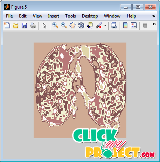
The major problems encountered are image segmentation and imperfect system response function. Image segmentation is defined as the process of classifying the voxels of an image into a set of distinct classes. The difficulty in PET image segmentation is compounded by the low spatial resolution and high noise characteristics of PET images. Despite the difficulties and known limitations, several image segmentation approaches have been proposed and used in the clinical setting including thresholding, edge detection, region growing, clustering, stochastic models, deformable models, classifiers and several other approaches. The strategies followed for validation and comparative assessment of various PET segmentation approaches are described. Future opportunities and the current challenges facing the adoption of PET-guided delineation of target volumes and its role in basic and clinical research are also addressed. A small lung lesion is only 2 or 3 voxels in diameter and of marginal contrast. It could easily be missed by human observers. Our method aims to pro vide radiologists with a map of potential lesions for decision so that diagnostic efficiency can be improved. It utilises both PET and CT images. The CT image provides a lung mask, to which lesion detection is confined, whereas the PET image provides distribution of glucose metabolism, according to which lung lesions are detected. Experimental results show that respiratory motion correction significantly increases the success of lesion detection, especially for small lesions, and most of the lung lesions can be detected by our method. The method can serve as a useful computer-aided image analysing tool to help radiologists read images and find malignant lung lesions.
Only logged in customers who have purchased this product may leave a review.


Reviews
There are no reviews yet.